


CT SCAN DEVICE
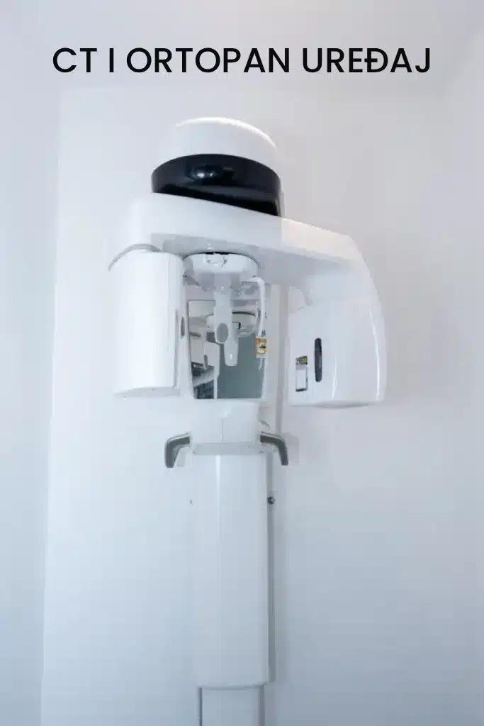
The CT device available at modern dental practices is essential today, both for implant diagnostics and for the overall assessment of dental conditions. It provides a three-dimensional image of the teeth, displaying anatomical structures, bone condition, and existing infections even more precisely than a panoramic X-ray.
PANORAMIC X-RAY (ORTHOPANTOMOGRAM)
A panoramic X-ray (orthopantomogram) is an image of both jaws that shows all teeth and surrounding anatomical structures. It is one of the main diagnostic methods and is an essential part of the initial patient examination when providing the most detailed and accurate treatment assessment.
At our dental clinic in Zagreb, Dental Estetic Studio, we have the latest panoramic X-ray technology, allowing patients to quickly and safely receive their scan during the first examination. The imaging process takes only a few seconds, and everything can be completed in one place. The digital image is provided on a CD, allowing you to store it and use it whenever needed, while we permanently keep it connected to your patient record.
The main advantage of this new-generation panoramic X-ray device is significantly lower radiation exposure compared to older systems, which is extremely important for patient safety.
Such imaging provides us with a more detailed view of all anatomical structures and allows us to establish a more accurate diagnosis.
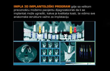
A panoramic X-ray is essential in all areas of dentistry, and starting treatment without it is extremely difficult. The latest developments in dentistry, especially in oral surgery, allow us to perform advanced bone analysis using panoramic imaging combined with specialized software. This enables us to analyze bone density, measure distances from the sinuses, determine the position of nerves, enlarge anatomical structures, and precisely plan future implant placement.
RVG – DIGITAL RADIOGRAPHY
RVG – digital radiovisiography enables imaging of individual teeth and provides the most detailed view of a tooth with significant magnification. This allows early detection of infections and assessment of bone condition. This type of imaging reduces patient radiation exposure compared to traditional X-ray methods.
Using an intraoral sensor, an image is captured and displayed on a computer monitor, where every detail can be carefully analyzed and evaluated.
BLOG POSTS YOU MAY FIND INTERESTING








REQUEST ADDITIONAL INFORMATION, CONSULT WITH US OR REQUEST A QUOTE
AND
WE WILL GET BACK TO YOU AS SOON AS POSSIBLE
Call us at +385 1 4847 432 or send us a message via the inquiry form.
DEŽMANOVA 5, ZAGREB, CROATIA
info@ordinacijaknego.hr